Greetings from the director
The Human Brain Research Center (HBRC) was established in 2000 to clarify the functional anatomy of the brain. We primarily employ non-invasive brain measurement techniques, primarily on humans, to investigate the neural correlates of the human mind. We also aim to understand the pathomechanisms of mental and neurological diseases and provide support for neurosurgical procedures.
Currently, our research is anchored in three core areas: advanced neuroimaging studies using a 7 Tesla MRI (Brain Function Imaging Field), clinical applications of Magnetoencephalography (MEG) for epilepsy treatment in collaboration with the hospital (Clinical Neurophysiology Field), and innovative research in Neurorehabilitation.
Now, we envision expanding our contributions beyond our own research activity. We plan to strengthen our capacity as a foundational facility supporting academic and clinical research, both within and beyond our institution. As a first and significant step, we participated in the establishment of the Center for Innovative Systems Neurotherapeutics (CiSNeuro) within Kyoto University Hospital. Our 7 Tesla MRI and MEG equipment are open for collaborative research. Researchers from other academic institutions, as well as corporations, are invited to utilize these resources. Please feel free to contact us for more information.
Members
Click here for the members of our lab.
Introduction to our research activities
We are engaged in the research of the structure of the human and animal brain, as well as the diagnosis and treatment of diseases that occur in the nervous system including the brain. We are also committed to the development of tools to investigate the brain. We aim to "understand the brain with the brain" by combining the following methods:
- Brain Function Imaging
- Clinical Neurophysiology
- Neurological Recovery and Regeneration Medicine
Brain Function Imaging Field
- Members: Naoya Oishi, Dinh Ha Duy Thuy, Shin-ichi Urayama, Hirotaka Onoe, Thai Akasaka
Ultra-high field MRI (Magnetic Resonance Imaging, Siemens MAGNETOM 7T)
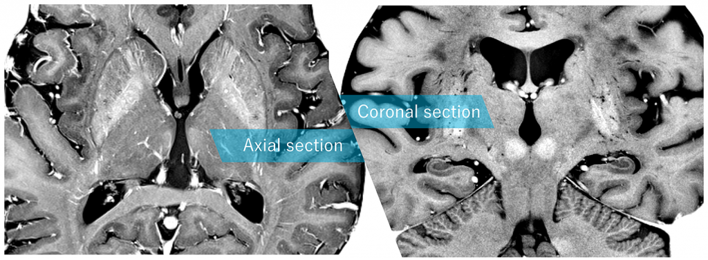
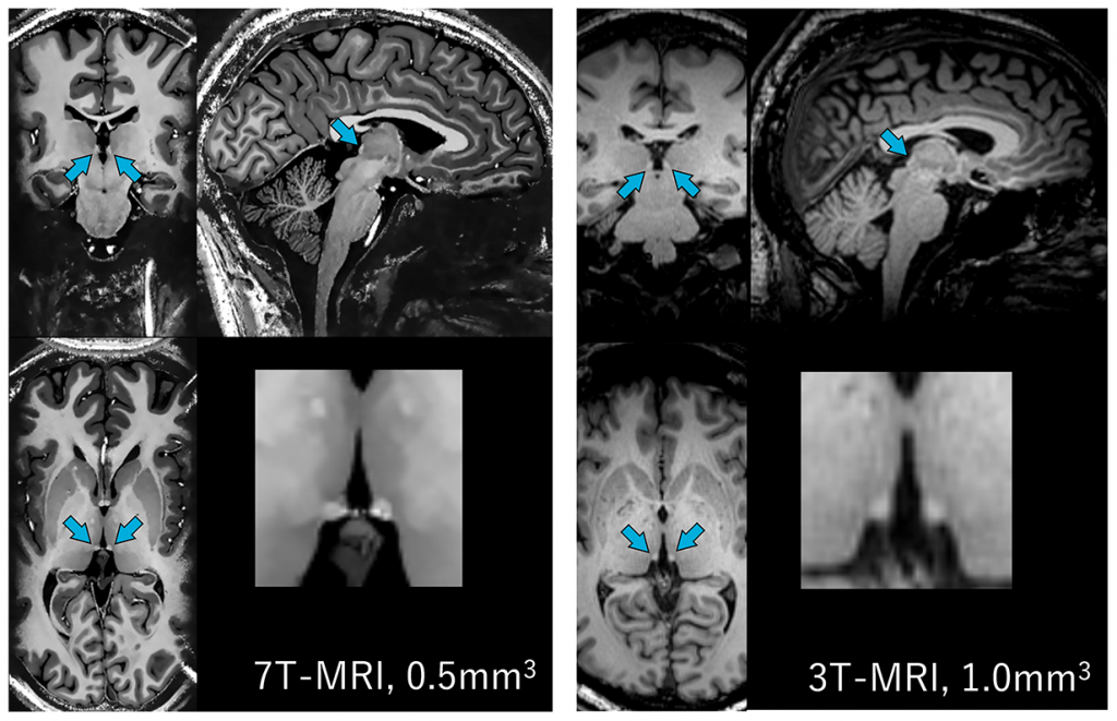
We installed an ultra-high field 7-tesla MRI (Siemens MAGNETOM 7T) in 2015 and utilizes its high signal strength and contrast to measure high-resolution and high-precision brain structural and functional images.

- Research using structural images
- Cerebral cortex volume, cortical thickness
- Observation of white matter fibers using diffusion tensor imaging (DTI)
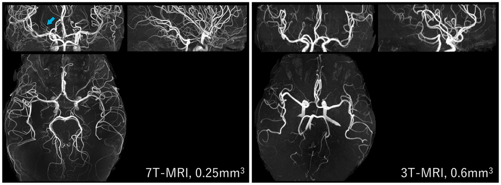
- Cerebral angiography with high spatial resolution
- Quantitative magnetic susceptibility imaging (QSM)
- Functional imaging (Functional MRI, fMRI)
- Functional brain mapping during task
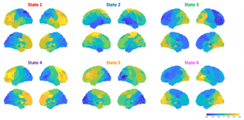
- Resting-state fMRI and functional connectivity
- Research on imaging methods
- High-speed imaging methods
In humans, we are conducting research not only in healthy subjects, but also in neurological and psychiatric disorders. We also study monkeys and various brain specimens.
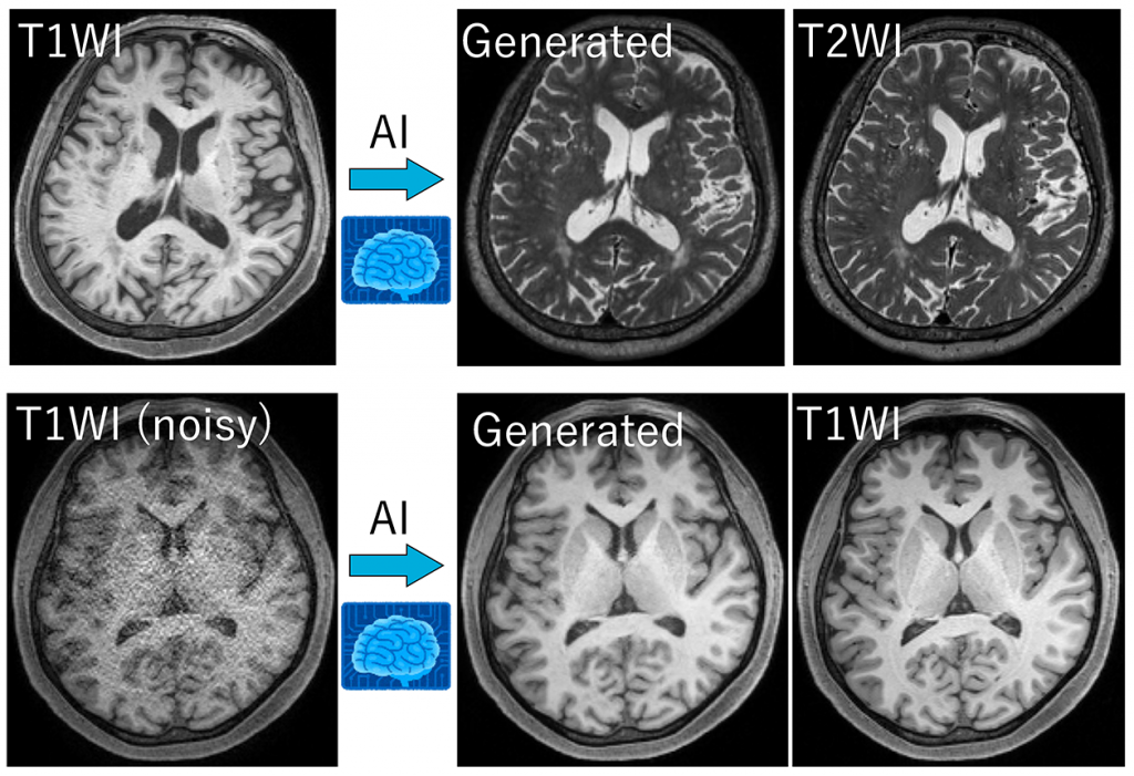
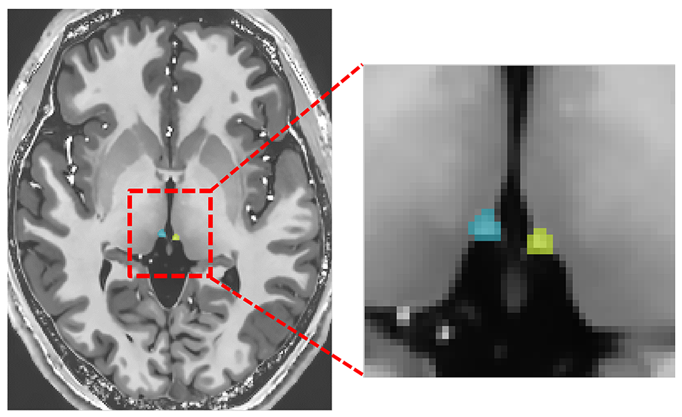
Research on Applications of Machine Learning and Deep Learning (Oishi N.)
We are conducting various researches using machine learning and deep learning in MRI. In addition to the development of essential technologies for image analysis, such as image-to-image translation, high-precision denoising, and accurate automatic identification of small brain structures, we are also conducting research in collaboration with many other researchers, with a view to clinical applications, such as elucidating pathological conditions in neurological and psychiatric disorders and developing prediction methods for therapeutic intervention.
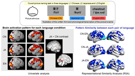
Language Network and Brain Function (Thuy DHD)
Using both task-based and resting-state functional MRI, we investigate the neural mechanisms of language processing and how the brain represents and organizes linguistic information in multilingual populations. Task-based paradigms, such as word generation, phonological and semantic association tasks, are used to probe controlled lexical retrieval and language production. In parallel, resting-state fMRI characterizes the intrinsic language network by capturing spontaneous neural activity and functional connectivity in the absence of explicit tasks. By integrating these approaches, we examine language representations at both regional and network levels, and how they are shaped by factors such as multilingual experience, age of acquisition, and language proficiency. This framework provides insights into the neural basis of language and its plasticity, with potential clinical applications.
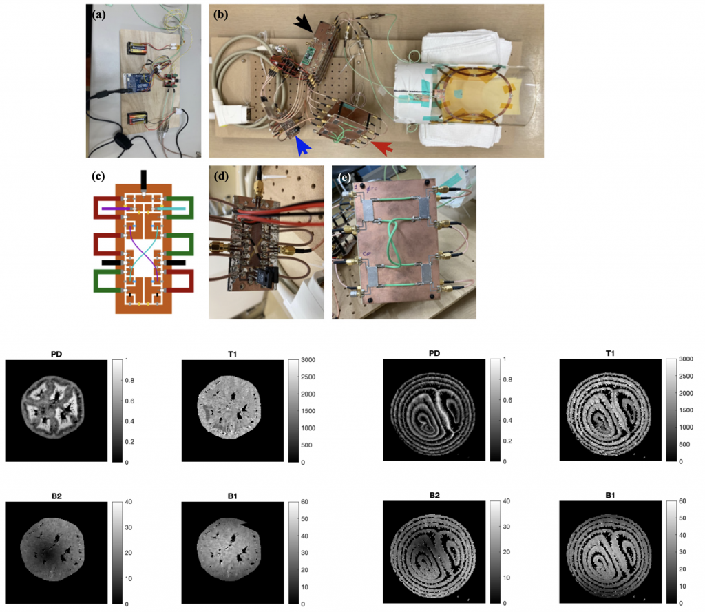
Development of RF coils for 7T-MRI (Urayama S)
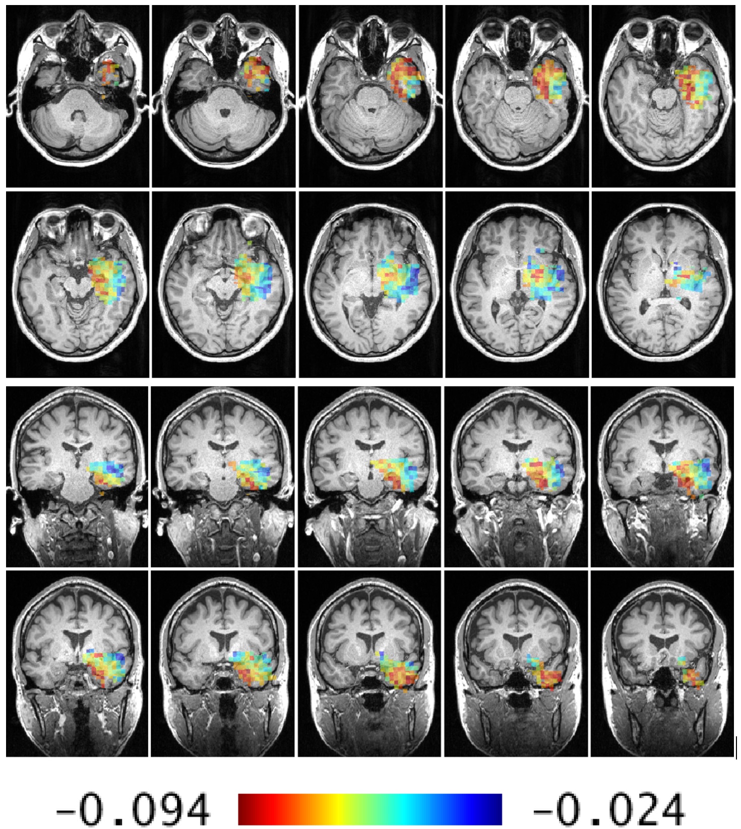
Depiction of cerebral aneurysm pulsations with ultra-high temporal resolution 3D-cine MRI (Akasaka T)
The fatality rates for ruptured intracranial aneurysms range between 35-50%, with up to half of those who survive experiencing long-term disabilities. Recently, localized irregular pulsations of cerebral aneurysms were discovered to be a risk factor for rupture. In this study, we utilize the high signal-to-noise ratio of 7T MRI and apply a technique called GRASP, which combines golden-angle radial scanning and compressed sensing, to depict the arterial wall movements of cerebral arteries as a 3D-cine image at ultra-high frame rates of up to 80 frames per second. We call this method GRASP-MRA and aim to establish it as a novel, non-invasive method for assessing the risks of cerebral aneurysm rupture.
Left: GRASP-MRA (65 frames per second) image of the cerebral arteries, including a right internal carotid artery aneurysm (approximately 8mm in size)
Right: A color map with the quantified degrees of wall motion of the cerebral aneurysm
MRI Research in Non-Human Primates (Onoe H)
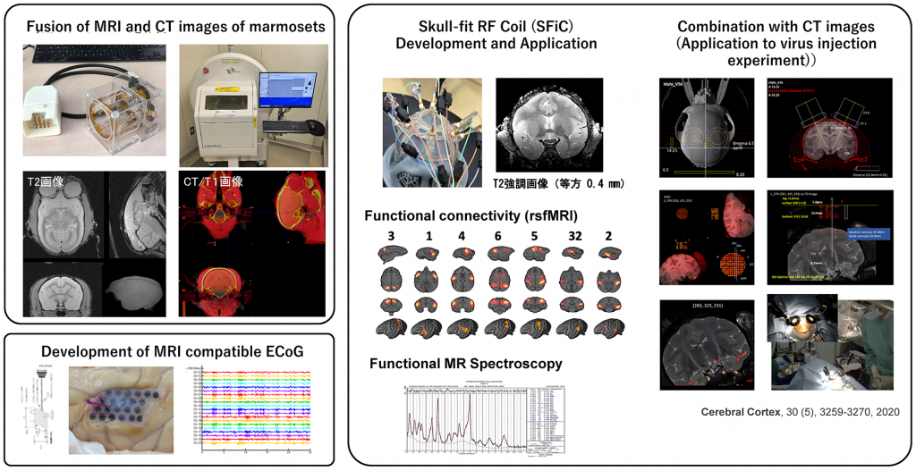
Understanding the structures and functions that support advanced cognitive functions in primates is crucial not only for elucidating the origins of human intelligence but also for understanding and developing treatments for refractory psychiatric and neurological disorders in humans. Common marmosets, due to their small size and smooth brain structure, facilitate challenging tasks such as simultaneous recording from extensive brain regions and Ca2+ imaging of cortical neuron population activity as well as circuit manipulations using viral vectors. This makes them promising for achieving neuroscience's critical goal of unraveling the circuitry underlying primate-specific higher cognitive functions. In recent years, experiments using non-human primates have rapidly advanced studies involving optogenetics and chemogenetics for circuit-selective manipulations. To enhance the precision of these studies, accurate knowledge of brain structures, the position of neuronal nuclei, and their relationship with the skull is necessary for precise targeting. We have established techniques for accurately fusing high-resolution CT images with 7T MRI and for precisely injecting viral vectors or drugs into targeted brain regions.
Higher brain functions in humans, especially higher cognitive functions, evolved in primate-developed systems. Analyzing brain functions using macaques, which are close relatives of humans, is crucial for understanding these functions and for advancing measurement techniques in brain function. We have successfully developed Skull Fit RF Coil (SFiC), based on individual cranial CT images using 3D printing technology, which provides significantly higher signal-to-noise ratios compared to conventional cylindrical RF coils, enabling high-quality resting-state functional MRI and Magnetic Resonance Spectroscopy (MRS) measurements.
2) Development of MRI-Compatible ECoG
Epidural or subdural cortical electrodes (ECoG) allow functional mapping of cortical activities with high spatiotemporal resolution. While MRI is commonly used for brain imaging in diagnosing brain disorders, embedding ECoG electrodes poses risks such as artifacts and tissue heating around metal electrodes. Therefore, in collaboration with materials engineers, we are developing highly compatible ECoG using carbon electrodes.
Clinical Neurophysiology Field
- Members: Madoka Matsumoto, Ryuta Aoki, Masao Matsuhashi
Magnetoencephalography (MEG)
The center introduced a 122-channel frontal MEG (Neuromag 122) in 1994, upgraded to its 306-channel successor (Vectorview) in 2004, and moved the MEG to the central clinical building of the hospital in 2021 to promote clinical research using MEG. We are developing and demonstrating more accurate diagnostic methods for various central nervous system and psychiatric disorders, including epilepsy. In addition, we are also focusing on basic research by conducting neurobiological studies on brain functions such as decision making, language, and memory in healthy individuals.
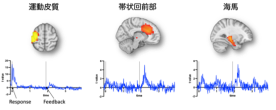
Mental health research based on neural dynamics (Matsumoto, Aoki)
Magnetoencephalography (MEG) is a method of non-invasively measuring the magnetic field generated by neural activity in the brain from the scalp. It is well known that representation for "self" in the brain by the neural network centering on the prefrontal cortex is involved in mental health, however it remains unknown how the "self" is represented by the dynamics of the neural network (neural dynamics) including the prefrontal cortex, and what kind of abnormalities in the neural dynamics are observed when the "self" is altered. We are engaged in mental health research by measuring brain functions using state-of-the-art analytical techniques such as neural decoding and state estimation of neural network.
Diagnosis of epilepsy and functional brain mapping using MEG(Matsuhashi)
We are developing and demonstrating more accurate diagnostic methods while using them in the clinical diagnosis of epilepsy and various other central nervous system disorders.
Link:Department of Epilepsy, Movement Disorders and Physiology
Visualization of the epileptogenic focus by Temporal Spread Imaging (TSI)
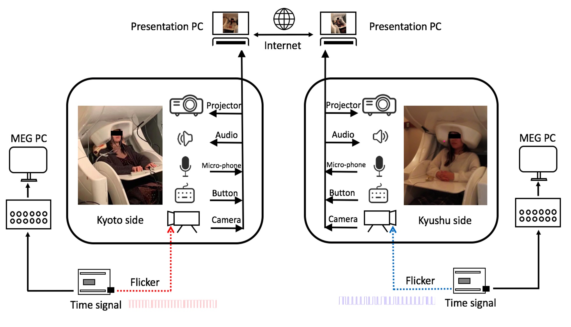
MEG hyperscanning via internet video connection with Kyushu University
Neurological Recovery and Regeneration Medicine Field
- Members: Satoko Koganemaru, Atsushi Shima, Kazuki Tanaka, Masako Yamada
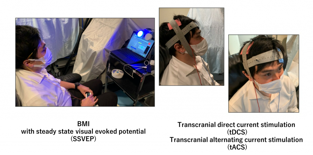
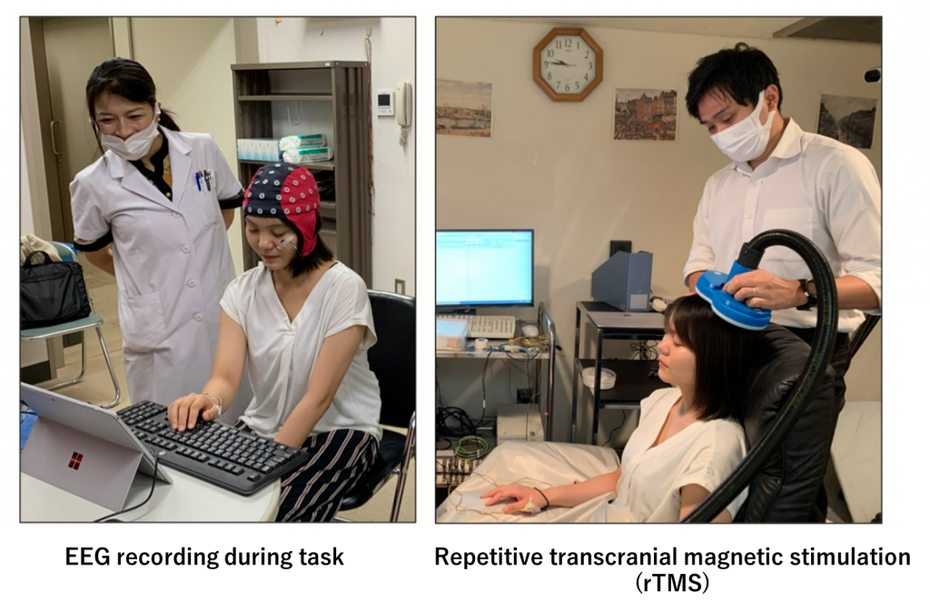
We are conducting state-of-the-art research aimed at efficient functional recovery by inducing brain plasticity through non-invasive brain stimulation (NIBS) combined with rehabilitation tasks. Our NIBS techniques are transcranial magnetic stimulation (TMS), transcranial direct current stimulation (tDCS), transcranial alternating current stimulation (tACS), transcranial static magnetic field stimulation (tSMS) and so on. We are also conducting research on Brain-Machine Interfaces (BMI) and robot rehabilitation, and further development and verification of those systems. In addition to developing new neuromodulation technologies for neurological disorders, we are conducting imaging research to clarify the neural basis of function recovery using magnetoencephalography (MEG), electroencephalography (EEG), and MRI measurements. Our research target is patients with neurological diseases (stroke, Parkinson's disease, dementia, etc.) and healthy individuals.
Publications
Click here for recent publications.
Human Brain Research Center
Graduate School of Medicine, Kyoto University
Director  Takashi Hanakawa (concurrent)
Telephone  +81-75-751-3695
FAX      +81-75-751-3202
email    hbrcoffice@kuhp.kyoto-u.ac.jp
URL.       http://hbrc.kuhp.kyoto-u.ac.jp/
Address   Third Clinical Research Bldg
           54 Shogoin-kawahara-cho, Sakyo-ku, Kyoto 606-8507, Japan
Nearest Stations
 - Kyodai Hospital (Kyodai Hospital Circular Route Bus HOOP)
 - Konoe-dori (Kyoto City Bus)
 - Jingu-Marutamachi Station (Keihan Train)